CareerPath for Imaging Scientists
During 2023 and 2024, the Global Bioimaging Career Paths for Imaging Scientists working group collected information on the challenges experienced by personnel in core facilities. Drawing on this data, the group has written an International Recommendation paper and, subsequently, a Journal of Microscopy Article on “Recognising the importance and impact of Imaging Scientists: Global guidelines for establishing career paths within core facilities”. The group aims to offer a global perspective on the challenges faced by core facility scientists and propose recommendations for nurturing their professional advancement, drawing insights from successful case studies.
🌐 Your Voice Matters! Share Your Challenges!
We would like to ascertain if you, as a member of this community, agree with our recommendations and if you can help us identify areas of concern that are relative to your institute, university, or facility and are not already represented or included. Our Top 5 Challenge Survey can be completed online here.
Our goal is to increase international submissions to strengthen representation and diversity, ensuring the broadest possible engagement with the topic. The survey will be open until the end of the year (31.12.2024). We plan to resurvey the community periodically to assess changes over time and determine the impact of our working group's efforts and published recommendations on the challenges Imaging Scientists face.
📧 Stay Connected, Stay Informed!
Ready to join this transformative journey? REGISTER HERE to receive ongoing updates on the TOP 5 landscape results.
GBI's Spotlight Seminar Series
University-Industry Collaboration: moving beyond simply providing services
19th Dec (5-6 pm CET) & 20th Dec (8-9 am CET)
Read more...
University-Industry Collaboration: moving beyond simply providing services
Each iteration of this monthly seminar series will feature speakers from the different Global BioImaging partner networks or other community members advancing career paths for Imaging Scientists working in core facilities. The seminar will be split between presentations and community discussions around the presented approaches, challenges and solutions.
To allow for broad attendance from the global imaging community, the seminars will be held twice on the same topic – one aimed at the Eastern hemisphere (8-9 am CEST, 3 pm JST, 4 pm AEST) and one for the Western hemisphere (5-6 pm CEST, 12 pm UYT/BRT/ART, 11 am EDT, 9 am CST, 8 am PDT). It will always take place on the last Wednesday/Thursday of the month. Watch all previous GBIs on the YouTube channel!
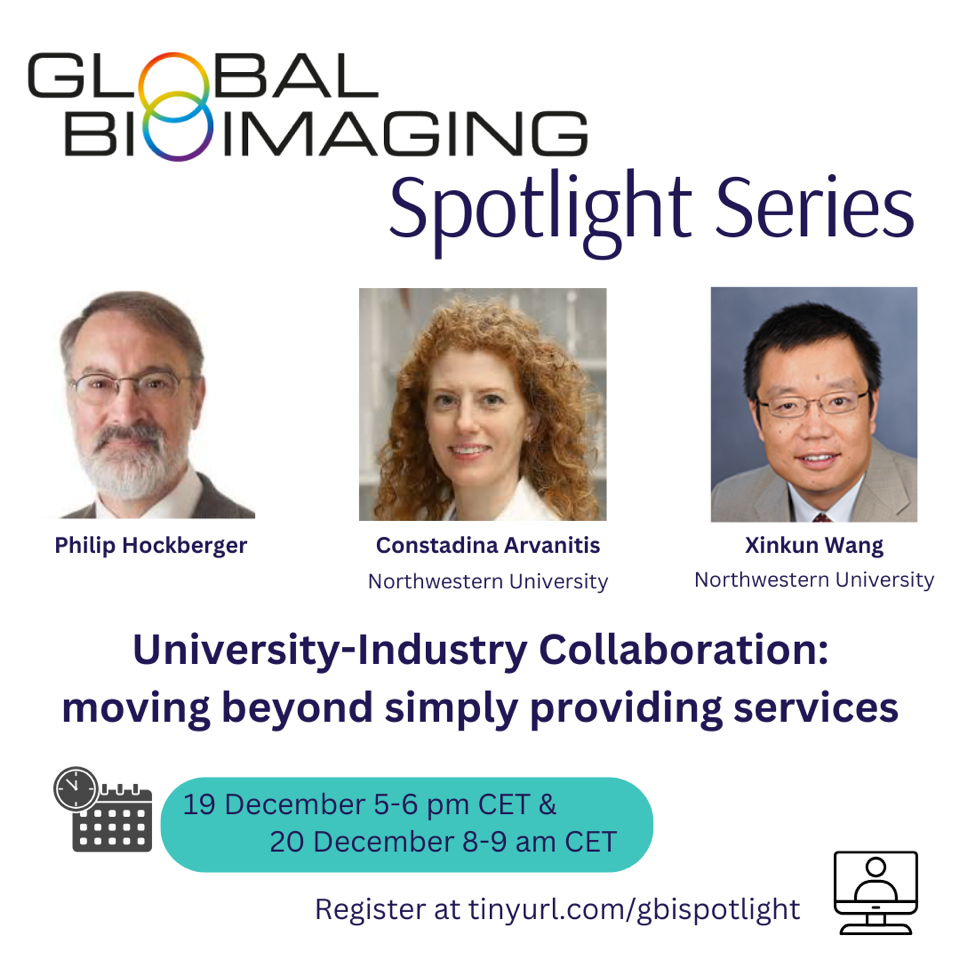
Philip Hockberger, Constadina Arvanitis and Xinkun Wang will explore diverse models of collaboration between core facilities and industry, showcasing how these partnerships can drive growth and innovation. From engaging with manufacturers and technology providers to offering services to industry users, such collaborations can expand core facilities’ reach, attract new user groups, and boost revenue. Successful partnerships require understanding industry needs, meeting specific requirements, and learning to "speak the industry language." Drawing from their experiences at the Northwestern University, the speakers will share how these interactions were built, the pathways to success, and the career impacts of working closely with industry.
The Global BioImaging Spotlights are open for all to attend – register at www.tinyurl.com/gbispotlight
From Trainee to Trainer: Building Networks and Opportunities in Bioimaging
28th Nov (5-6 pm CET) & 29th Nov (8-9 am CET)
Read more...
From Trainee to Trainer: Building Networks and Opportunities in Bioimaging
Each iteration of this monthly seminar series will feature speakers from the different Global BioImaging partner networks or other community members advancing career paths for Imaging Scientists working in core facilities. The seminar will be split between presentations and community discussions around the presented approaches, challenges and solutions.
To allow for broad attendance from the global imaging community, the seminars will be held twice on the same topic – one aimed at the Eastern hemisphere (8-9 am CEST, 3 pm JST, 4 pm AEST) and one for the Western hemisphere (5-6 pm CEST, 12 pm UYT/BRT/ART, 11 am EDT, 9 am CST, 8 am PDT). It will always take place on the last Wednesday/Thursday of the month. Watch all previous GBIs on the YouTube channel!
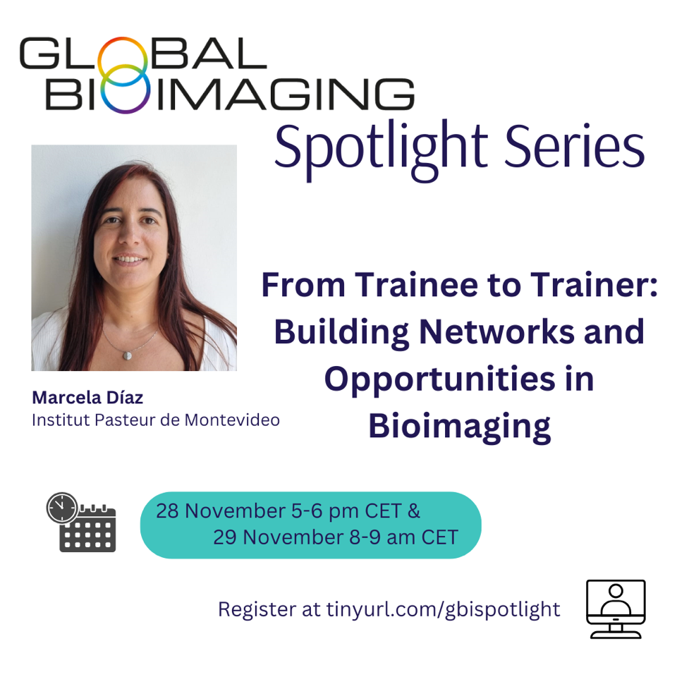 Marcela Diaz will highlight the importance of teaching and training in shaping a successful core facility role. Starting as a junior facility member participating in a "train-the-trainer" course, Marcela has advanced to leading such courses herself and teaching imaging methods to researchers and other imaging scientists. In this talk, she will discuss how developing both scientific and pedagogical skills to effectively teach and train users is critical for core facility staff - not only as a tool for career growth but also as a means to make a broader impact by sharing imaging knowledge.
The Global BioImaging Spotlights are open for all to attend – register at www.tinyurl.com/gbispotlight
(Why) Is an MBA useful for a core facility microscopy manager?
August 29th (8-9 am CEST & 5-6 pm CEST)
Read more...
(Why) Is an MBA useful for a core facility microscopy manager?
Each iteration of this monthly seminar series will feature speakers from the different Global BioImaging partner networks or other community members advancing career paths for Imaging Scientists working in core facilities. The seminar will be split between presentations and community discussions around the presented approaches, challenges and solutions.
To allow for broad attendance from the global imaging community, the seminars will be held twice on the same topic – one aimed at the Eastern hemisphere (8-9 am CEST, 3 pm JST, 4 pm AEST) and one for the Western hemisphere (5-6 pm CEST, 12 pm UYT/BRT/ART, 11 am EDT, 9 am CST, 8 am PDT). It will always take place on the last Wednesday/Thursday of the month. Watch all previous GBIs on the YouTube channel!
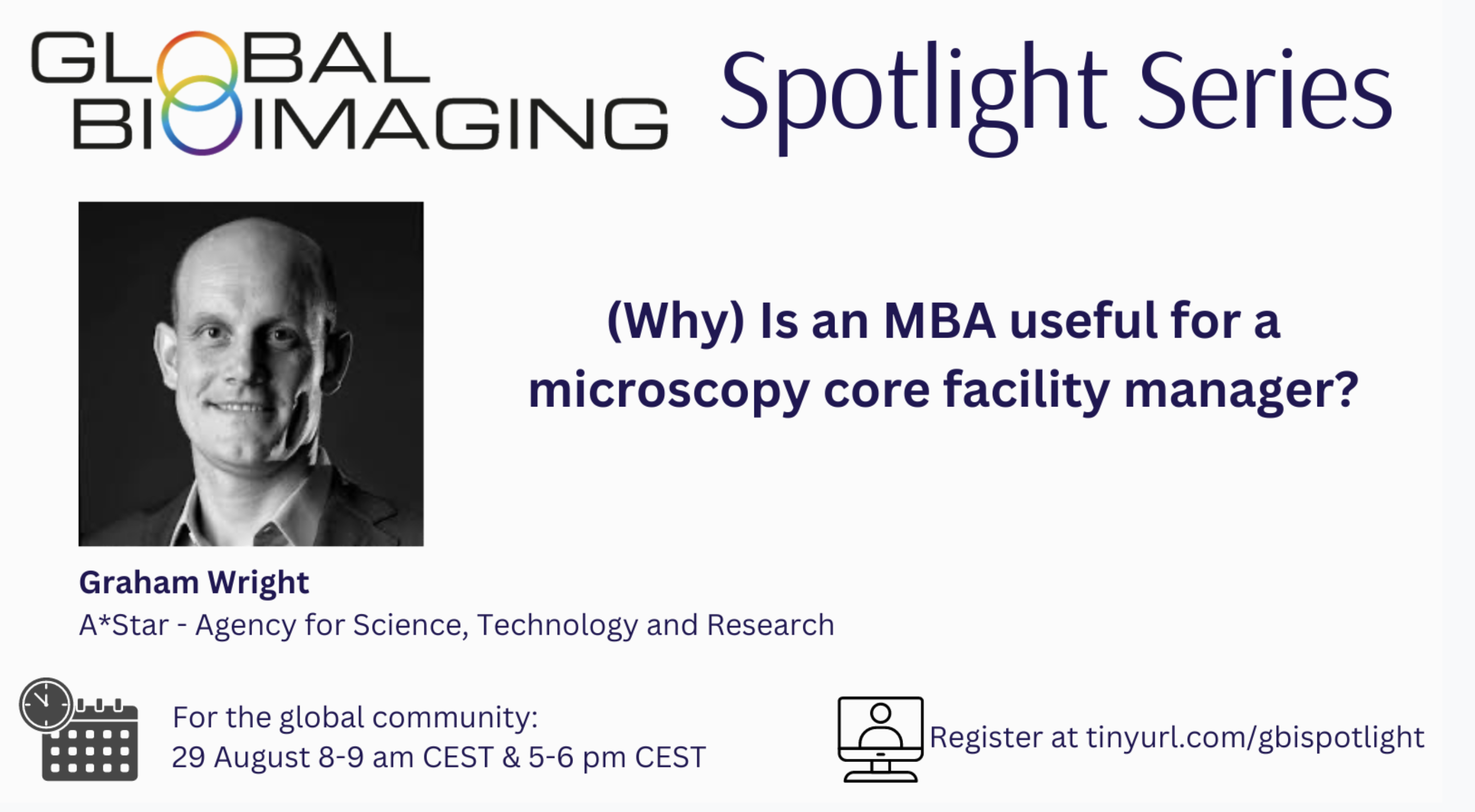
Graham Wright exemplifies how pursuing a traditional MBA, such as his two-year part-time program at Warwick Business School, can significantly enhance the management and coordination of research facilities. His experience highlights the value of integrating business skills like marketing, strategic thinking, and financial planning into the professional development of Imaging Scientists, advocating for increased institutional support for such educational opportunities.
The Global BioImaging Spotlights are open for all to attend – register at www.tinyurl.com/gbispotlight
Charting an imaging science career path in Africa
27th June (5-6 pm CEST) & 28th June (8-9 am CEST)
Read more...
Charting an imaging science career path in Africa
Each iteration of this monthly seminar series will feature speakers from the different Global BioImaging partner networks or other community members advancing career paths for Imaging Scientists working in core facilities. The seminar will be split between presentations and community discussions around the presented approaches, challenges and solutions.
To allow for broad attendance from the global imaging community, the seminars will be held twice on the same topic – one aimed at the Eastern hemisphere (8-9 am CEST, 3 pm JST, 4 pm AEST) and one for the Western hemisphere (5-6 pm CEST, 12 pm UYT/BRT/ART, 11 am EDT, 9 am CST, 8 am PDT). It will always take place on the last Wednesday/Thursday of the month. Watch all previous GBIs on the YouTube channel!

The Africa Microscopy Initiative (AMI) aims to address unequal access to advanced microscopy in Africa. It offers an all-expense-covered open-access microscopy center, educational programs, and an instrument distribution scheme. AMI recognizes that effective microscopy capacity-building in Africa relies on technology access, dissemination, and education. Fully funded by the Chan-Zuckerberg Initiative and the Gates Foundation, AMI also facilitates the transfer of pre-owned microscopes to African institutions with demonstrated need. It provides international fellowships and mentorship opportunities to African microscopists, expanding trained personnel for microscopy teaching. The first call-for-proposal for the AMI Imaging Centre received 63 proposals from 18 countries. AMI's impact extends to global access programs like the Advanced Imaging Center (AIC) at HHMI Janelia Research Campus, which has seen increased submissions from Africa. AMI's long-term sustainability depends on African funding agencies' support and raising awareness within the scientific community, especially in Africa, where the concept of open-access research and training-the-trainers may be unfamiliar.
The Global BioImaging Spotlights are open for all to attend – register at www.tinyurl.com/gbispotlight